Article
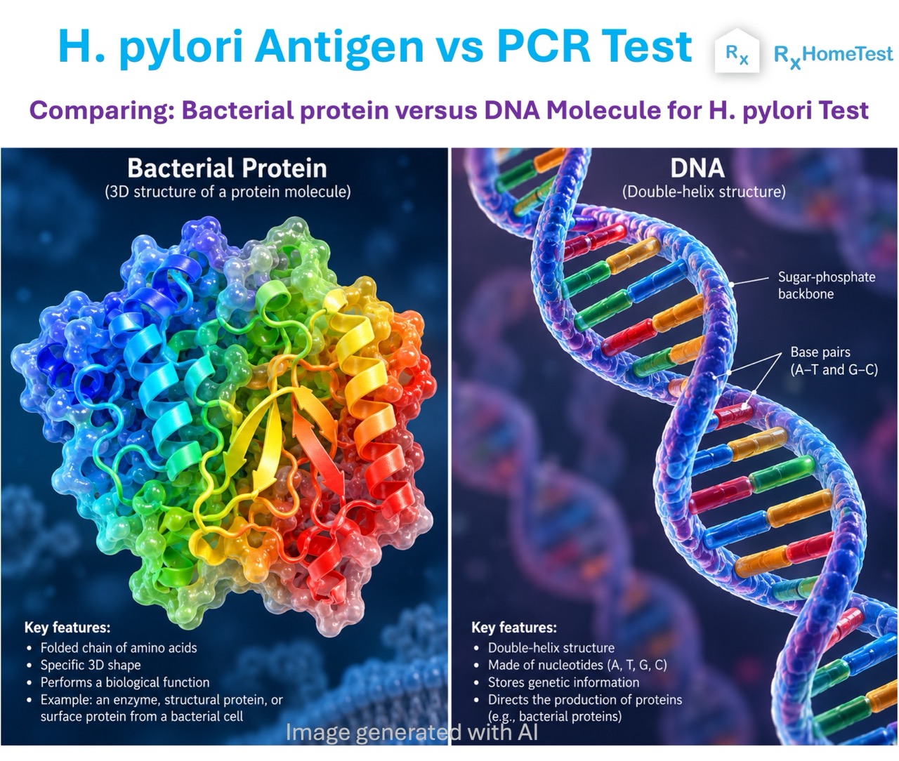
Here we explain the difference between two different kinds of helicobacter pylori tests.
When it comes to checking for H. pylori, there are several options.
Understanding the different kinds of H. pylori tests is important for choosing the right diagnosis and treatment for persistent stomach pain, acid reflux, or suspected ulcers.
While both of these non-invasive methods use stool samples, they detect fundamentally different parts of the bacteria and offer very distinct insights for treatment.
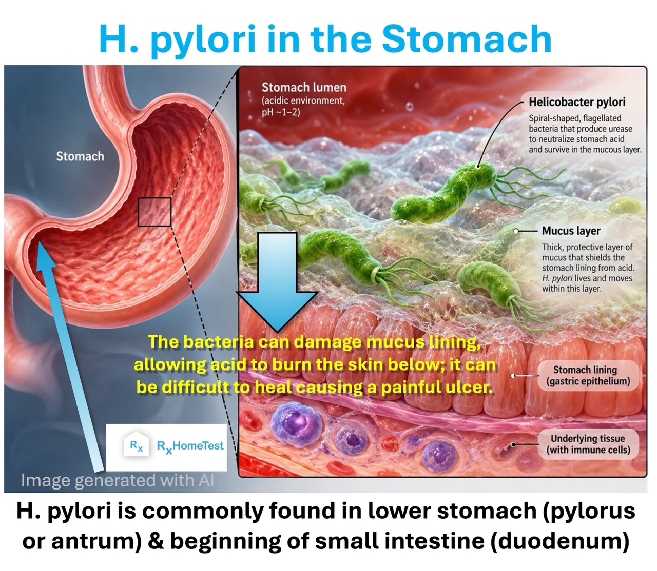
Figure: An illustration of Helicobacter pylori bacteria inhabiting the protective mucosal lining of the human stomach.
Helicobacter pylori (H. pylori) is a spiral-shaped bacterium that colonizes the human stomach lining.
According to National Institutes of Health (NIH), it affects an estimated 50% of the world's population. It's especially common in less developed countries.
This persistent pathogen is a primary cause of chronic gastritis, peptic ulcer disease, and gastric adenocarcinoma (stomach cancer).
While many infected individuals show no symptoms for decades, others experience severe digestive symptoms that require timely and accurate clinical diagnosis.
Historically, diagnosing an active H. pylori infection required invasive procedures such as an upper gastrointestinal endoscopy paired with a gastric mucosal biopsy.
Fortunately, advances in non-invasive tests have made it possible to detect the bacterium reliably using stool and breath samples.
Modern molecular and immunological technologies permit patients to collect samples conveniently, whether through their gastroenterologist's office or via reliable at-home health testing kits.
Among non-invasive diagnostic options, the stool antigen test (SAT) and the polymerase chain reaction (PCR) stool test are two of the most widely utilized laboratory assays.
Although both tests utilize stool as the biological specimen, they rely on entirely different biochemical principles, offer differing levels of analytical sensitivity, and deliver distinct diagnostic data.
Deciding between them depends on whether an individual needs a straightforward initial screen or a comprehensive molecular profile that includes antibiotic resistance detection.
H. pylori has evolved special survival mechanisms that allow it to survive in the highly acidic environment of the stomach.
By producing large quantities of the enzyme urease, the bacterium converts stomach urea into carbon dioxide and ammonia.
This chemical conversion neutralizes gastric acid around the bacterium's immediate vicinity, creating a neutral microenvironment that shields it from digestive enzymes.
Furthermore, the bacterium uses its flagella to corkscrew through the protective gastric mucus layer, adhering tightly to the underlying epithelial cells.
Over time, this colonization triggers localized inflammation and damages the mucosal barrier (as shown in the image below).
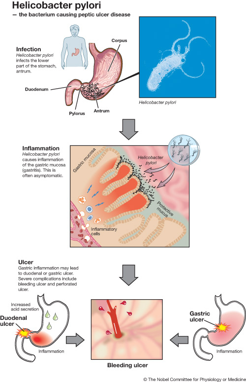
Figure: A close-up sketch showing how the bacteria colonizes the stomach and causes an ulcer in the gastric mucosa.
This process can lead to erosion of the mucous lining, duodenal ulcers, and recurrent functional dyspepsia with symptoms of burning epigastric pain, early satiety, bloating, and nausea.
Prolonged colonization can stimulate chronic atrophic gastritis, which the Centers for Disease Control and Prevention (CDC) and the World Health Organization classify as a known precursor to gastric cancer.
Given the long-term cancer and inflammatory risks from untreated infection, an accurate diagnosis is essential.
False-negative results can leave an active infection untreated, allowing mucosal damage to progress.
Conversely, false-positive results can trigger unnecessary regimens of heavy multi-drug antibiotic therapy, disturbing the gut microbiome, provoking adverse drug reactions, and exacerbating global antimicrobial resistance.
The stool antigen test (SAT) is an established, non-invasive immunoassay designed to detect the presence of H. pylori surface proteins (antigens) shed directly into the digestive tract.
When the bacteria colonize the gastric mucosa, their outer membrane proteins and cellular fragments continuously slough off, travel through the intestines, and are excreted in the feces.
Modern clinical laboratories perform this test using enzyme-linked immunosorbent assays (ELISA) or lateral flow immunochromatographic assays.
During the assay, antibodies that are specifically calibrated to bind to H. pylori antigens are exposed to the processed stool sample.
If the target bacterial proteins are present, they bind to these antibodies, producing a measurable colorimetric or fluorescent signal that confirms the presence of active bacterial shedding.
Stool antigen assays are broadly categorized into polyclonal and monoclonal varieties.
Monoclonal antibody tests, which use identical immune cells cloned from a single parent cell, offer markedly higher specificity and sensitivity than older polyclonal preparations.
Because the test detects actual bacterial antigens rather than host antibodies, a positive result reflects an active, ongoing colonization rather than a past, resolved infection.
Polymerase chain reaction (PCR) testing is an advanced molecular diagnostic approach that targets the genetic material of H. pylori.
Rather than seeking out intact bacterial proteins, a real-time PCR assay extracts total microbial DNA from a stool sample and exponentially replicates specific, highly conserved gene sequences unique to the H. pylori genome.
Common genetic targets in H. pylori PCR assays include genes such as ureA, glmM (ureC), and the 23S rRNA gene.
During the automated thermal cycling process, laboratory reagents including DNA polymerase, primers, and fluorescent probes bind specifically to target fragments of bacterial DNA.
Each cycle doubles the quantity of target DNA, enabling clinical instruments to detect even microscopic amounts of bacterial genetic material that traditional immunoassays might miss.
Because real-time quantitative PCR (qPCR) monitors DNA amplification as it happens, it provides an exceptionally clear threshold for identifying active infections.
Molecular testing can detect low bacterial loads, atypical strains, or degraded bacterial particles that might lack intact surface antigens, rendering it an indispensable tool in modern gastrointestinal clinical microbiology.
The fundamental distinction between an antigen test and a PCR test lies in the biological analyte each method targets.
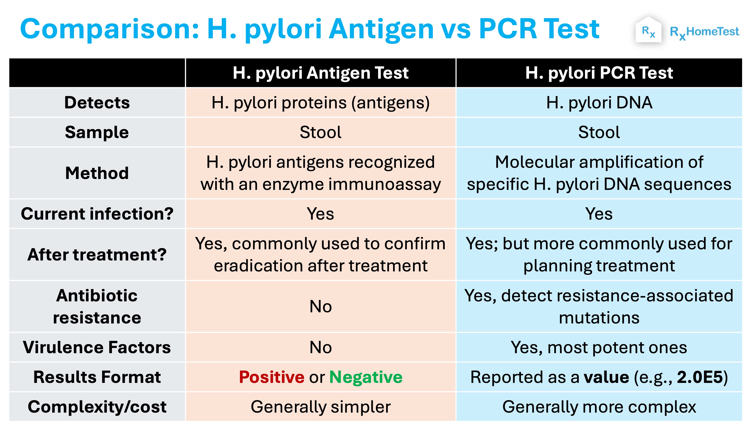
Table: Comparing antigen test vs a PCR DNA test.
Stool antigen tests assess phenotypic expression, specifically, the structural outer membrane proteins produced by viable, intact bacteria.
In contrast, PCR tests evaluate genotypic markers by identifying the precise nucleic acid sequences that make up the bacterial chromosome.
This technical difference matters for diagnostic sensitivity and sample viability.
Bacterial proteins can degrade rapidly as they pass through the small intestine and colon, where they are exposed to proteolytic enzymes, digestive bile salts, and competing colonic microflora.
If stool transit time is delayed, or if a sample is stored at room temperature for an extended period, the structural integrity of these antigens can break down, reducing the likelihood of detection by immunoassay antibodies.
DNA molecules, by comparison, are chemically more resilient than complex quaternary protein structures.
Real-time PCR protocols are engineered to amplify short genetic fragments, meaning that even if the bacterial cell has lysed and its proteins have broken down during digestion, its diagnostic DNA sequences often remain intact.
This makes PCR testing inherently less vulnerable to sample degradation caused by delayed transit or transit-related temperature fluctuations.
The accuracy of laboratory tests is measured using two parameters: sensitivity (the ability to correctly identify someone with an infection) and specificity (the ability to correctly identify someone who doesn't have one).
Both stool antigen and stool PCR assays demonstrate exceptional diagnostic metrics, but minor differences exist depending on clinical context.
According to clinical consensus guidelines published by the Mayo Clinic and the American College of Gastroenterology (ACG), high-quality monoclonal stool antigen tests typically achieve a sensitivity of 88% to 95% and a specificity of 92% to 97%.
These figures make monoclonal antigen assays reliable first-line tools for standard screening.
However, if someone has a low bacterial load or loose, watery stool that dilutes antigen concentrations, the sensitivity of the antigen test can fall noticeably.
Stool PCR testing generally demonstrates clinical sensitivity ranging from 93% to 98% and specificity exceeding 98%.
Because amplification generates billions of copies from a single DNA strand, the limit of detection (LoD) for PCR is substantially lower than that of immunoassays.
Consequently, PCR is less prone to false-negative results in borderline cases or mild colonization, delivering greater diagnostic certainty when symptoms persist despite negative preliminary screenings.
The rise of antibiotic resistance in H. pylori represents a pressing challenge in clinical gastroenterology.
The World Health Organization has designated clarithromycin-resistant H. pylori as a high-priority pathogen for research and development.
Traditional first-line triple therapy—comprising a proton pump inhibitor, clarithromycin, and amoxicillin—now fails in up to 30% of patients in regions with elevated macrolide resistance.
This is where PCR testing offers a major diagnostic benefit that antigen tests cannot match.
While an antigen test provides only a binary "positive" or "negative" result, multiplex real-time PCR can simultaneously analyze the 23S rRNA gene of H. pylori for point mutations (such as A2142G, A2143G, and A2142C). This allows designing a treatment regimen specific to your antibiotic resistance and severity of your infection.
These point mutations are directly responsible for conferring bacterial resistance to clarithromycin.
Some advanced molecular panels also screen for mutations in the gyrA gene, which dictate resistance to fluoroquinolones like levofloxacin.
By identifying these genetic resistance mutations straight from a non-invasive stool sample, PCR testing allows clinicians to practice precision medicine.
Rather than relying on trial-and-error empiric therapy, healthcare providers can tailor the person's antibiotic regimen to the specific bacterial strain present, significantly increasing first-line eradication rates and protecting overall gut wellness.
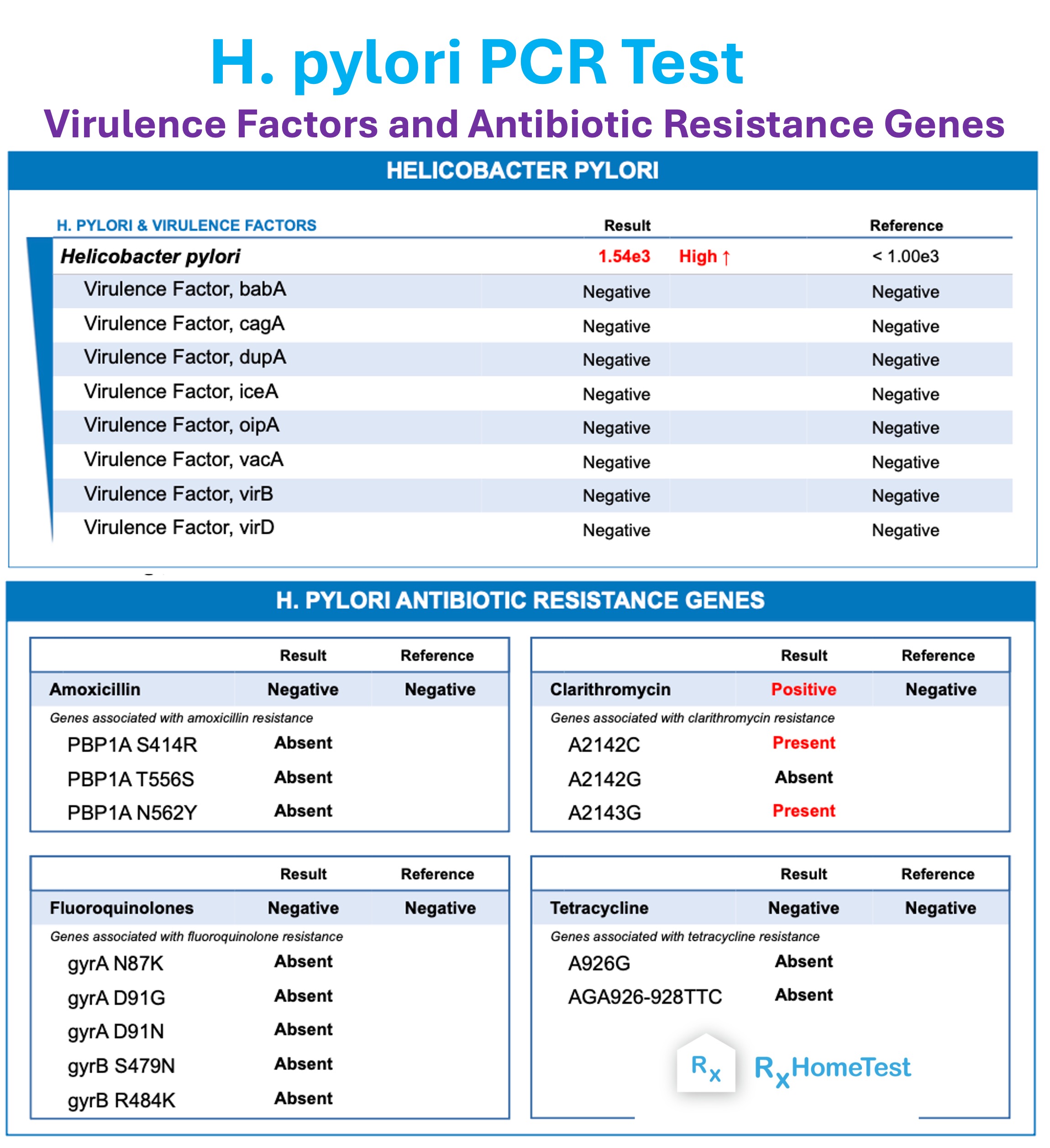
Figure: A PCR test has the added advantage of insights about the more infectious virulence factors and the patient's antibiotic resistance, allowing their healthcare provider to design a highly targeted treatment regimin.
Both antigen and PCR assays are subject to pharmacological interference, and medication-induced false negatives represent a common pitfall in clinical practice.
The primary culprits behind inaccurate test results are proton pump inhibitors (PPIs), bismuth subsalicylate compounds, and antibacterial agents.
Proton pump inhibitors, such as omeprazole, esomeprazole, and pantoprazole, suppress gastric acid production, raising the pH inside the stomach.
While this provides symptom relief, it also inhibits H. pylori metabolism and forces the bacteria to transition from their active spiral morphology into an inactive, dormant coccoid form.
In this state, bacterial replication slows down, gastric colonization density drops, and antigen shedding decreases dramatically, which frequently leads to false-negative stool antigen results.
To ensure diagnostic accuracy, clinical guidelines emphasize strict pre-test protocols:
Proton Pump Inhibitors (PPIs): Discontinue use for at least 14 days prior to sample collection. Patients can often transition temporarily to H2-receptor antagonists (like famotidine) or standard antacids under clinical guidance.
Antibiotics and Bismuth Compounds: Discontinue use for at least 28 to 30 days prior to stool collection. These medications directly diminish bacterial density throughout the gastrointestinal tract.
While PCR tests are marginally more tolerant of lower bacterial concentrations due to their amplified sensitivity, taking these medications within the restricted window can still suppress bacterial DNA levels below the assay's limit of detection.
Diagnosing an initial H. pylori infection and confirming its successful eradication following medical therapy require distinct clinical approaches.
A patient experiencing unresolved gastrointestinal complaints may benefit from either a monoclonal antigen test or a stool PCR test as an initial screening tool, depending on regional resistance trends and individual medical history.
However, when performing a "test of cure" to verify that an infection has been eradicated, the timing and choice of diagnostic method become critical.
Standard professional protocols dictate that post-treatment confirmation testing must occur no earlier than four to six weeks following the completion of antibiotic therapy.
Testing too soon can produce misleading results regardless of the test chosen.
In this post-treatment setting, monoclonal stool antigen testing is widely favored because dead, non-viable bacteria are eliminated from the digestive tract within days of completing treatment.
Therefore, the presence of surface antigens four weeks later strongly indicates treatment failure and active bacterial persistence.
PCR, while highly sensitive, can occasionally detect residual, non-viable bacterial DNA fragments for several weeks after the organisms have been eradicated, which carries a small potential risk of a transient false-positive outcome if performed too early.
Modern at-home laboratory collection protocols have removed traditional barriers associated with clinical stool sample submission.
Previously, patients had to coordinate laboratory drop-offs during strict business hours with temperature-sensitive collection containers.
Modern at-home testing services supply clear, standardized instructions alongside specialized collection devices and preservation buffers.
Patients collecting specimens at home should follow these essential steps to protect sample integrity:
Avoid Toilet Water Contamination: Utilize the provided collection paper or collection hammock to capture the sample cleanly, avoiding any contact with toilet bowl water or chemical cleansers, which can degrade proteins and inhibit PCR enzymes.
Sample Preservation: Immediately place the stool aliquot into the designated transport tube. Many modern PCR and antigen collection vials contain specialized transport media that stabilize proteins and nucleic acids during transit.
Timely Dispatch: Seal the sample securely in the biohazard bag and return it via the provided prepaid courier packaging as directed by the kit instructions.
For individuals looking to understand broad digestive issues alongside targeted pathogen detection, pairing specific diagnostic tests with a comprehensive gut microbiome test can offer broader insights into bowel regularities, microbial diversity, and intestinal inflammation.
When choosing between an H. pylori stool antigen test and an H. pylori PCR test, several practical parameters must be evaluated, including diagnostic goals, availability, laboratory turnaround times, and out-of-pocket costs.
Stool antigen testing is widely available in nearly every commercial and hospital laboratory globally.
Due to its streamlined immunoassay methodology, it is typically the more cost-effective option and boasts rapid laboratory processing times, often returning results within 24 to 48 hours.
For straightforward initial screenings in communities with low known antibiotic resistance, the monoclonal stool antigen test remains an accessible, highly reliable choice.
Stool PCR testing involves sophisticated molecular processing and specialized laboratory equipment, making it somewhat less universally available than antigen tests. But it provides much more information.
However, the higher investment is often justified for patients with recurrent symptoms, individuals who have experienced previous treatment failures, or those who need molecular antibiotic resistance screening before initiating a complex quadruple-therapy regimen.
Choosing PCR equips the clinician with actionable genetic data that can shorten the overall duration of illness and prevent ineffective antibiotic cycles.
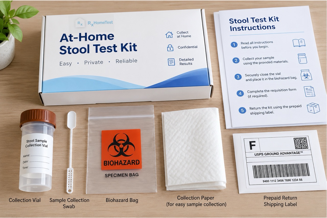
Figure: An example of supplies included in an at-home H. pylori test kit.
Actually there are at least two more options:
The antibody test checks for antibodies produced by the body against the Helicobacter pylori bacteria (that's why the name).
But once someone is exposed, the test will almost always be positive (since the body will have the antibodies circulating in the blood).
Therefore, it may not be able to test current infection.
It is often used to test family members to confirm if others are also infected and whether they need treatment.
You normally draw blood in a vial or with finger prick and mail it to the lab to check for H. pylori antibodies.
A breath test includes two small pouches; you breathe into them, seal and mail to the lab.
First pouch collects natural levels of carbon dioxide in your breath.
After drinking a small amount of solution (that has a different carbon molecule), you collect a second sample.
It looks for the presence of urease enzyme, produced by the bacteria (urease breaks down urea).
Urease is not produced by our body, and a positive result means presence of the bacteria in the second sample.
In order to survive the harsh acidic environment of the stomach, H. pylori breaks down urea and creates a protective sack around itself.
Both the H. pylori stool antigen test and the stool PCR test serve vital, complementary roles in modern gastrointestinal medicine.
While the stool antigen test remains an accessible, cost-effective, and accurate tool for routine initial detection and post-treatment verification, real-time PCR provides elevated analytical sensitivity and indispensable genetic information regarding antibiotic resistance mutations.
Selecting the right test requires evaluating your personal clinical picture, previous exposure to antibiotics, history of treatment failures, and the timing of medication washouts.
Discussing these factors with a qualified medical provider ensures that your diagnostic strategy provides definitive answers, leading to targeted treatment, long-term gastric mucosal healing, and lasting gastrointestinal comfort.
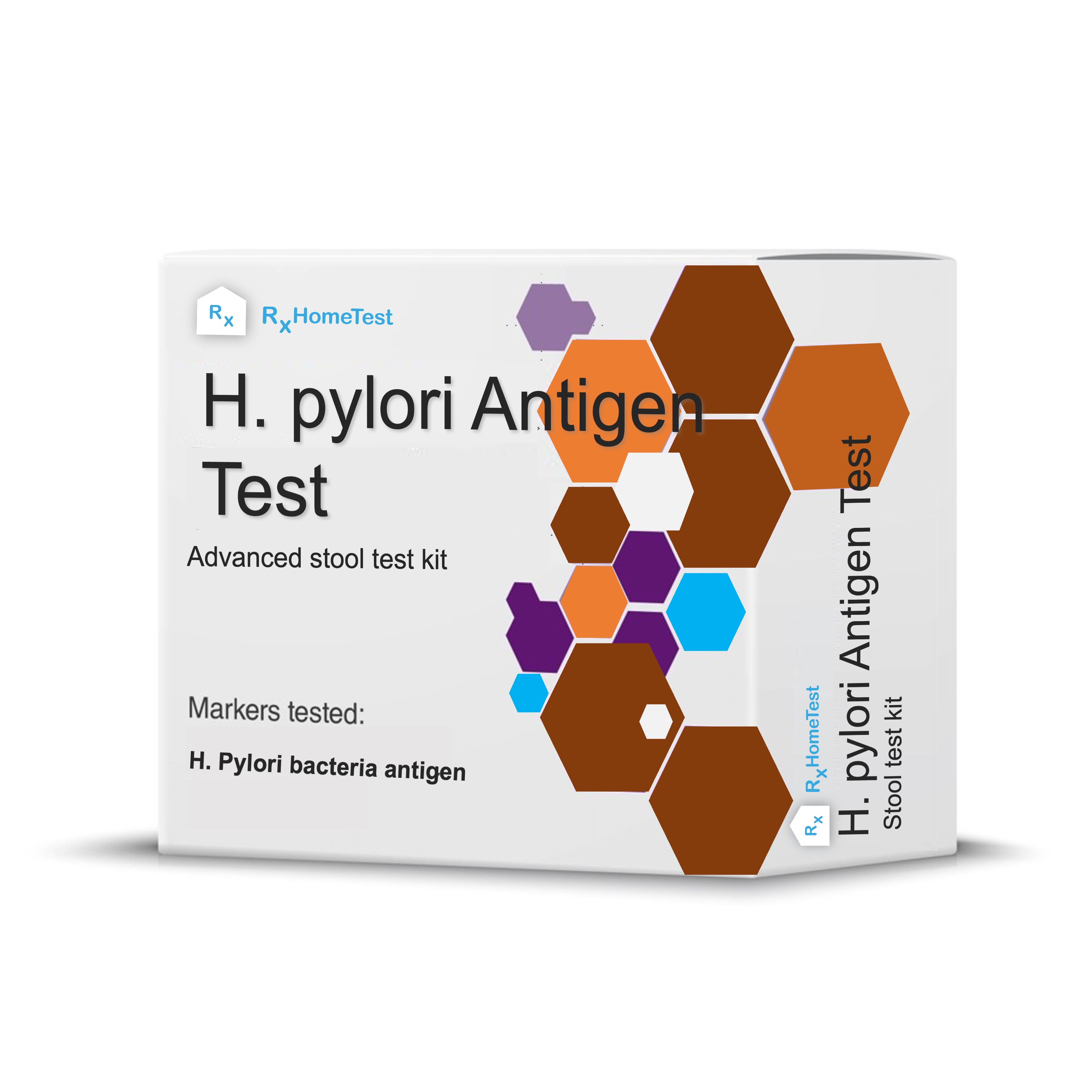
Order an at-home H. pylori antigen test.
Stomach Ulcers and H. pylori – take a deep dive into the cause of ulcers.
H. pylori and Acidity - Key Facts – find out how the bacteria contribute to acidity.
Are At-Home Tests FSA/HSA Eligible – read before you order a test kit.
1. National Institutes of Health (NIH): National Institute of Diabetes and Digestive and Kidney Diseases (NIDDK). Helicobacter pylori and Peptic Ulcers. Available from: nih.gov
2. Centers for Disease Control and Prevention (CDC): Helicobacter pylori: Fact Sheet for Healthcare Providers. Available from: cdc.gov
3. Chey, W. D., Leontiadis, G. I., Howden, C. W., & Moss, S. F. (2017): ACG Clinical Guideline: Treatment of Helicobacter pylori Infection. The American Journal of Gastroenterology, 112(2), 212-239.
4. Mayo Clinic Laboratories: Helicobacter pylori Diagnostic Testing Algorithms and Stool Molecular PCR Evaluations. Available from: mayocliniclabs.com
5. Malfertheiner, P., et al. (2022): Management of Helicobacter pylori infection—the Maastricht VI/Florence consensus report. Gut, 71(9), 1724-1762.
